A Brief History of Magnetic Resonance Imaging
Damadian identifies T1/T2 differences between cancer and normal. He was seeking an MR signal difference in an important disease (cancer) that would prove his idea of an MR body scanner was a goal worth pursuing. Submits paper. Founds Fonar Corporation.
Sir Peter Mansfield – realized that adding a magnetic field gradient could allow scientists to look at the atomic structure of a chemical.
July 3, 1977 – The first MRI body scan is performed on a
human using an MRI machine developed by American doctors
Raymond Damadian, Larry Minkoff and Michael Goldsmith.

Stages in the development of MRI
“…advances can be roughly classified into hardware (eg, magnets, gradients, radiofrequency [RF] coils, RF transmitter and receiver, MR imaging–compatible biopsy device’s) and imaging techniques (eg, pulse sequences, parallel imaging, and so forth). Hybrid systems, such as MR/positron emission tomography (PET), combine the superb anatomic and functional imaging capabilities of MR imaging with the unsurpassed capability of PET to demonstrate tissue metabolism. Supported by the improvements in hardware, advances in pulse sequence design and image reconstruction techniques have spurred dramatic improvements in imaging speed and the capability for studying tissue function. In this historical review, the history of MR imaging technology and developing research and clinical applications, as seen through the pages of Radiology, will be considered.“
https://pubs.rsna.org/doi/full/10.1148/radiol.14140706
MRI’s have evolved and are used to help diagnose a wide variety of medical conditions, including:
- Abnormalities of the brain/spinal cord
- Injuries and abnormalities of the joints
- Liver and abdominal diseases
- Uterine abnormalities
- Heart problems
- Tumors and cysts
- Abnormalities of the brain and spinal cord
Diffusion Tensor Imaging (DTI) is an MRI-based neuroimaging technique which makes it possible to estimate the location, orientation, and anisotropy of the brain’s white matter tracts.
Pioneers in the development of MRI
•1944: Rabi Physics (Measured magnetic moment of nucleus)
•1952: Felix Bloch and Edward Mills Purcell Physics (Basic science of NMR phenomenon)
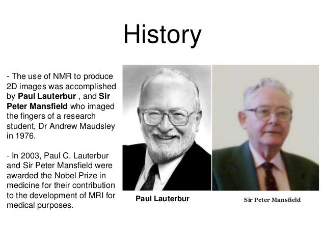
In 1973, Paul Lauterbur (State University of New York) described a new imaging technique that he termed Zeugmatography. By utilizing gradients in the magnetic field, this technique was able to produce a two-dimensional image (back-projection).
In 1975, Richard Ernst introduced 2D NMR using phase and frequency encoding.
By 1977, Dr. Raymond Damadian completed construction of the first whole-body MRI scanner, which he dubbed “Indomitable.”
Sir Peter Mansfield made the famous breakthrough at the University of Nottingham in 1977, which led to MRI, or Magnetic Resonance Imaging, being used in hospitals all over the world. He shared a Nobel Prize in 2003, with Paul C. Lauterbur, for his contribution to physiology and medicine.
•1991: Richard Ernst Chemistry (High-resolution pulsed FT-NMR)
•2002: Kurt Wüthrich Chemistry (3D molecular structure in solution by NMR)
•2003: Paul Lauterbur & Peter Mansfield

Worldwide, thirty-five 7-Teslas have been installed, and purely for research – none have entered the clinical routine.
[computerhistory.org]
ROADMAPS TO THE FUTURE:
ULTRAHIGH FIELD NMR AND MRI: SCIENCE AT A CROSSROADS
University of California Berkeley
Major increases in field strengths will enable unprecedented resolution for human imaging.
• Low-gamma and quadrupolar nuclei MRI and MRS will become feasible at high fields
B0 and B1 field distortions at high MR proton frequencies will enable imaging the electrical properties of the brain, leading to hitherto untapped sources of contrast and information.
https://www.osti.gov/servlets/purl/1338047
REFERENCES:
[http://sumerdoc.blogspot.com]
Time line and Nobel prizes in MRI
https://pubs.rsna.org/doi/full/10.1148/radiol.14140706
https://blog.healthcare.oxinst.com/the-history-of-the-mri-from-the-first-mri-to-today/
https://www.thoughtco.com/magnetic-resonance-imaging-mri-1992133