A Brief History of the Computerized Tomography Scanner

[GE Healthcare]
1917 – Johann Radon showed that a function can be reconstructed from an infinite set of its projections using the ‘Radon Transformation‘
Dr. Shinji Takahashi – the first to advocate the basic concepts of CT in the 1950’s. It is clear that Professor Takahashi understood the principle of modern CT in the 1950s, long before Godfrey N. Hounsfield reported the development of the first CT images in 1972.
CT was independently discovered by Dr. Alan Cormack and British engineer Sir Godfrey Hounsfield, who were jointly awarded the Nobel Prize in 1979 for their instrumental efforts in medicine.
First CT scanners available 1976, originally dedicated to head imaging only.
the EMI Mark 1, could scan only the brain. It was displayed in November 1972 at the Radiological Society of North America’s (RSNA) annual meeting
First CT scanners available in 1976, were originally dedicated to head imaging only
Steps in the development of CT

CT became widely available by 1980.
However, it took days to construct a single image
It is claimed the CT Scanner was the “greatest legacy” of the Beatles; the massive profits from their record sales enabled EMI to fund scientific research.Steps in the Development of CT
- In the early 1900s an Italian radiologist named Alessandro Vallebona invented tomography which used radiographic film to see a single slice of the body. As conventional tomography evolved, it was still considered ineffective when it came to imaging soft tissues.
- Increased power and availability of computers in the 1960s sparked the research to create practical computational tomographic images.
- In 1967 Sir Godfrey Hounsfield invented the first CT scanner at EMI Central Research Laboratories using x-ray technology.
1903 – E.A.O. Pasche builds a collimator for suppressing scattered radiation.
1917 – Johann Radon showed that a function can be reconstructed from an infinite set of its projections using the ‘Radon Transformation‘
1927 – Neurologist Dr Moniz performs the first cerebral angiogram.
*The first clinical scan in the USA was performed at the Mayo Clinic in 1973.
1974 – A fourth-generation CT system was developed. Fourth-generation CT systems employed a stationary gantry
*1976 saw 17 companies offering scanners, with scan times down to 5 seconds in some cases.
*Development continued through the 1990s, with the introduction of spiral (continuous) scanning in the early 1990s and the development of multi-slice scanners, with 4-slice scanners and 0.5 second scan times being ‘state-of-the-art’ by the end of the century
Pioneers in the Development of CT
” At least ten people independently invented tomography over the period 1921 to 1934 with absolutely no idea that the others were working towards the same goals. “
Contributors to Tomography:
Allesandra Vallenona: Stratigraphy
Bernard Ziedses: Planigraphy
Z. des Plantes: Laminography
Andre Brocage: Zonography
1977 – The first whole-body computed tomograph from Siemens, the SOMATOM
Following SOMATOM, Siemens introduces an improved version of its head scanner, the SIRETOM 2000. A resolution four times higher than its predecessor, better image quality, as well as faster and more comfortable examinations are just some of the benefits of the new scanner.
In 1984, Siemens produces a further innovation: An entire CT system is installed in a trailer truck so that it can be transported to hospitals and practices that cannot afford their own CT.
A Journey through the History of Computed Tomography – Part Two: From Top to Toe with SOMATOM

A 512 X 512 inch image can now be constructed in less than a second.
Computed Tomohttp://www.theevolutionofimagingtechnology.com/andre-brocage/graphy: Prof. E. Lobel
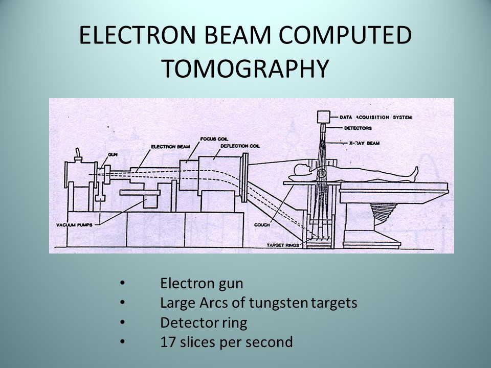
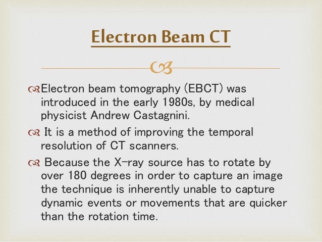
Electron Beam Tomography(EBCT) was introduced in the early1980s, by medical physicist Andrew Castagnini.
/
First Generation: Rotate/Translate, Pencil Beam
The first CT scanners were solely for head scans and used a rotate/translate system with a single x-ray beam called a pencil beam.
Second Generation: rotate/Translate, Narrow Fan Beam
The largest advantage associated with the second generation CT scanner was the substantial decrease in acquisition time compared to that of the first generation. Although the angle of the fan beam was not large and still required the linear movement of the x-ray tube and detectors at each projection angle, the amount of linear displacement required was dramatically reduced [3]
Third Generation: Rotate/Rotate, Wide Fan Beam
Translational motion, which was used in first- and second-generation scanners, was quite time consuming. At this stage in development, the main goal was to cut the acquisition time to less than 20 seconds so that the brain could be imaged more rapidly, but also so that physicians could image parts of the body other than the head [1].
Fourth Generation: Rotate/Stationary
Fourth generation scanners were developed specifically to alleviate the ring artifacts produced by the third generation. Specifically, the impossibility to have such a large array of rotating detector elements (>400) are perfectly synced and calibrated to one another. By removing the detectors from the rotating gantry and putting them in a stationary ring around the patient, detectors were able to maintain calibration [4]
Fifth Generation: Stationary/Stationary
Fifth generation CT scanners were developed specifically for use in cardiac tomography imaging. These scanners were often referred to as cine-CT scanners, or more commonly electron beam scanners [2]
Sixth Generation: Helical
A slip ring allows electricity to be passed to rotating components without needing stationary components [5]. Using a slip ring allowed the gantry to rotate continuously through all of the patient slices, therefore creating shorter scan times. This led to the development of the sixth generation CT scanner, also known as helical CT (or, less accurately, spiral CT) (Figure 6).
Seventh Generation: Multiple Detector Array
Unlike the pencil beam and fan beam, the cone beam does not pass through a narrow collimator. Therefore, the intensity of the initial x-ray beam is not as strongly reduced and hence can interact more efficiently and effectively with the detector array [1]
The Radon Transformation is widely applicable to tomography
Almost as soon as the x-ray was discovered it was realised that planar radiographs, being two-dimensional projections of three-dimensional structures, showed the organs and structures of interest shadowed by over- and under-lying structure, thus reducing their diagnostic efficacy. A brief history of tomography and CT

| Convolution |
| Convolution is an important mathematical technique in digital signal processing. Raw data undergo spatial filtration prior to back projection by combining two signals to form a third signal. Convolution is related to the input signal, the output signal, and the impulse response. This operation is mostly used together with Fourier transformations for CT signal and image processing. https://www.radiology-tip.com/serv1.php?type=db1&dbs=Convolution |

Alessandro Vallebona constructed equipment and published the first clinical body-section imaging material ever in 1930, but his method was not ideal. The first clinical material employing an ideal method was published by Bernhard Ziedses des Plantes in 1932. Methods for transverse axial tomography was independently described by William Watson in 1937, Jean Kieffer in 1938, and Shinji Takahashi in 1947.
https://www.ncbi.nlm.nih.gov/pubmed/11625126
Cone beam computed tomography (or CBCT, also referred to as C-arm CT, cone beam volume CT, or flat panel CT) is a medical imaging techniqueconsisting of X-ray computed tomography where the X-rays are divergent, forming a cone.[1]
Cone beam technology was first introduced in the European market in 1996 by QR s.r.l. (NewTom 9000) and into the US market in 2001.
A brief history of tomography and CT
Steve Webb
” At least ten people independently invented tomography over the period 1921 to 1934 with absolutely no idea that the others were working towards the same goals. ”
” A Frenchman Andre Edmond Marie Bocage patented the concepts of tomography in 1921. These described how by simultaneously moving the x-ray source and detector in a ynchronised motion, ablurred tomogram resulted. He never built equipment but the Dutchman Bernard Zeidses des Plantes did, writing up the work in a doctoral thesis in 1934 and some papers a year or two earlier. He had also begun his work in 1921. Zeidses des Plantes, bom in 1902, lived until 1993 and provided personal comment to me on these early developments when I prepared my book of the history. Two other Frenchman, Felix Portes and Maurice Chausse also patented tomography in 1921 and made workshop drawings of how to implement it. A German engineer Ernst Pohl patented apparatus in 1927. The French Canadian Bartelink patented in 1931 and the GermanGustave Grossmannpatented in 1934. Grossmann built the first commercial equipment via his company Siemens-Reiniger-Veifa GmbH and the “Grossmann tomograph” became the most widelyused, if expensive, equipment in the late 193O’s. A surviving example is in the London Science Museum. Meanwhile, in Italy, Alessandro Vallebonapublished the tomography concept in 1931 and built apparatus. “
https://inis.iaea.org/collection/NCLCollectionStore/_Public/28/028/28028148.pdf
By 1975, EMI were marketing a body scanner, the CT5000, the first of which was installed at Northwick Park Hospital in London. The first body scanner in the USA was installed at the Mallinkrodt Institute and had its first clinical use in October 1975. By this time scan time had been reduced to 20 seconds, for a 320 x 320 image matrix.

Roadmaps to the Future of CT Imaging:
Fusion Imaging: Fusion imaging is the technology which enables
superimposition of images from two dependent imaging modalities to produce an image which provides greater information.
Quantitative Imaging. Quantitative imaging is the “extraction of quantifiable features from medical images for the assessment of normal or the severity, degree of change, or status of a disease, injury, or chronic condition relative to normal”55.
ECONOMICS OF CT IMAGING:
In the present era with several options, ‘expected workload’ is definitely the key in selecting the right CT scanner. 4 or 8-detector MDCT system is often sufficient for low-resolution imaging; coronary angiography requires a 64- detector system. A
RADIATION CONCERNS IN CT IMAGING:
This issue has initiated a sort of ‘dose war’ in the CT technology and manufacturers now claim to generate the desired image quality at lower radiation dosage for a particular imaging study.
http://medind.nic.in/jav/t13/i1/javt13i1p35.pdf
References:
International Society for Computed Tomography
International Society for Computed Tomography
https://wiki.uiowa.edu/display/881886/CT+Historical+Time+Line
http://199.116.233.101/index.php/Generations_of_CT_Scanners
A Journey through the History of Computed Tomography – Part Two: From Top to Toe with SOMATOM
http://ctscannerinfo.blogspot.com History of the Computerized TomographyA brief history of CT i
mpactscan.org History of the Computerized Tomography
https://www.siemens-healthineers.com/computed-tomography/news/mso-the-history-of-ct-2.html